
Leyenda de las figuras:
Figura 1: Dos grandes manchas café con leche (MCL) lumbosacras, con coloración menos homogénea y más difusa en zona paravertebral.
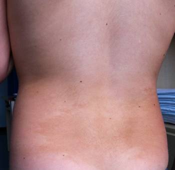
Figura 2: En el costado derecho, a nivel del tórax, se aprecia otra gran mancha café con leche (MCL) similar a las anteriores, pero de menor tamaño.
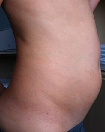
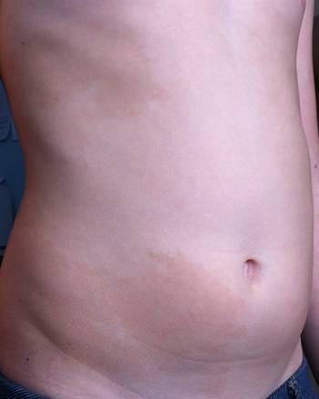
Figura 3: La mancha café con leche (MCL) mayor se delimita claramente a nivel de la línea media ventral, sin rebasarla.
Bibliografía
- Lacour JP. Taches café-au-lait. Ann Dermatol Venereol 1999 ;126 :749-54.
- Shah KN. The diagnostic and clinical significance of café-au-lait macules. Pediatr Clin North Am 2010; 57:1131-1153.
- Chateil JF et al. Phacomatoses chez l´enfant. Encycl Med Chir, Pediatrie, 4-092-B-10, 2000, 22p. Ed Elsevier SAS, París.
- Neurofibromatosis: conference statement. National Institutes of health consensus development conference. Arch Neurol 1988; 45:575-578.
- Hager CM, Cohen PR, Tschen JA. Segmental neurofibromatose: case reports and reciew. J Am Acad Dermatol 1997;37:867-69.
- Dumitrescu CE, Collins MT. McCune-Albright síndrome. Orphanet J Rare Dis 2008; 3:12-20
- Collins MT, Cantante RF, Eugster E. McCune-Albright syndrome and the extraskeletal manifestations of fibrous dysplasia.:http://www.ojrd.com/content/7/S1/S4.
- Hogelin M, Frieden IJ. Segmental Pigmentation disorder. Br J Dermatol 2010; 162:1337-1341.
- Listernick R, Mancini AJ, Charrow J. Segmental neurofibromatosis in chilhood. Am J Med Genet 2003; 121A:132-5.
- Metzker A, Morag C, Weitz R. Segmental Pigmentation disorder. Acta Derm Venereol 1983; 63:167-169.
- Happle R. Mosaicism Human Skin Understanding the Patterns and Mechanism. Arch Dermatol 1993; 129:1460-1470.