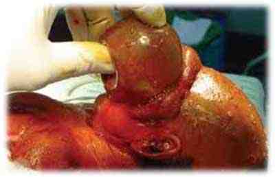
Figura 8. Se observa la cubierta de piel (señalado con la flecha roja) y meninges integras (señalado con la flecha verde)
Referencias bibliográficas
- Autores cubanos: Pediatría I, La Habana- Cuba, Ciencias Medicas, 2006: Parte VI Genética Médica, Cap. 29 Diagnóstico prenatal, Página 313.
- T.W. Sadler, Ph.D.: Lagman Embriología Medica con orientación clínica, 9na Edición, Buenos Aires- Argentina, Panamericana S.A., 2005: Cap. 7 Defectos congénitos y Diagnóstico prenatal, Pág. 170.
- Olney RS, Mulinare I: trends in neural tube defact prevalence, folic acid fortification, and vitamin supplement use. Semin Perinatol 26:277,2002
- http://www.cdc.gov/ncbddd/spanish/birthsdefects/encephalocele.html
- Holder-Espinasse M, Winter RM (2003). Type 1 Arnold–Chiari malformation and Noonan syndrome. A new diag-nostic feature? ClinDysmorphol 12: 275.
- http://www.scielo.org.pe/img/revistas/cimel/ulln2/a12fig04.html
- Sarnat HB, Menkes JH (2000). The new neuroembryology: how to construct a neural tube. J Child Neurol 15: 110–124.
- Camargo, Ulloa, Calvo y Lozano: radiología básica básica editorial Celsus, 2001
- Henrry, JB: El Labaoratorio y el diagnostico clínico. Editorial Marbàn 2005.
- Longman C, Mercuri E, Cowan F, et al. (2004). Antenatal and postnatal brain magnetic resonance imaging in mus-cle–eye–brain disease. Arch Neurol 61: 1301–1306.
- Williams LJ, et al: Prevalenceofspina bifida and anencephalyduringthe transition to mandatory folic acid fortification in the UnitedStates.Teratology 66:33, 2002.